Suite 5, Level 1
66 Pacific Hwy
ST LEONARDS NSW 2065
66 Pacific Hwy
ST LEONARDS NSW 2065
Fax: 02-8412 0060


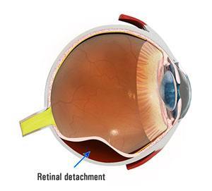
The retina is a nerve layer located at the back of the eye. This layer senses light and sends images to your brain. Like in a camera, the lens in front of the eye focuses light onto the retina. So the retina is like the film that lines the back of a camera.
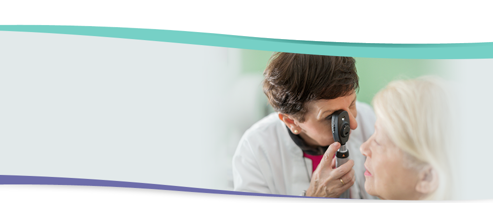
A retinal detachment occurs when the retina is pulled away from its normal position. The retina does not function when it is detached, so there is loss of vision from that detached part. A retinal detachment is a serious problem that almost always progresses to cause blindness unless it is treated. Your ophthalmologist can diagnose retinal detachment during an eye examination where the pupils of your eyes are dilated (enlarged) with drops.
The vitreous is a clear gel that fills the middle portion of the eye. As we get older, the vitreous may pull away from its attachment to the retina in one or more places. This may cause a retinal tear. Fluid may then pass through the retinal tear lifting the retina off the back of the eye, like wallpaper peeling off a wall.
Conditions which increases the chance of a retinal detachment
These following symptoms may indicate the presence of a retinal detachment.
Flashing lights are caused when the vitreous gel pulls on the retina. What you see may look like flashing lights or lightning streaks. You may have experienced this same sensation if you have ever been hit in the eye and seen “stars”.
Flashes of light in the eye are a serious warning symptom. Even though the flashes of light can appear off and on for several weeks or months, and the older we grow the more common it is to experience flashes, nevertheless they are important danger signs. If you notice a sudden appearance of light flashes, you should visit your ophthalmologist immediately.
Some people experience flashes of light that appear as jagged lines or “heat waves” in both eyes, often lasting for 10-20 minutes. These flashes are caused by spasm of the blood vessels in the brain which are called migraines. If a headache follows the flashes, this is called a Migraine Headache. If these flashes occur without a headache then these flashes are called Ophthalmic Migraine.
You may sometimes see small specks or clouds moving in your field of vision. These are called floaters. You can often see them when looking at a plain back ground, like a blank wall or blue sky. Floaters are tiny clumps of gel or cells inside the vitreous. While these objects look like they are in front of your eye, they are actually floating inside. What you see are the shadows they cast on the retina. Floaters can differ in shapes:
When people reach middle age, the vitreous gel may alter thickness or shrink thus forming clumps or strands inside the eye. These symptoms do not always mean a retinal detachment is present; however, you should see your ophthalmologist as soon as possible.
Most retinal tears need to be treated with laser surgery or cryotherapy (freezing), which seals the retina to the back of the eye. These treatments may cause discomfort and may require local anaesthetic injection or even general anaesthetic (going to sleep for surgery). However treatment of small tears may be performed in your ophthalmologist’s office.
There are several ways to fix a retinal detachment. The decision of which type of surgery depends upon the characteristics of your detachment. In each of the following methods your ophthalmologist will locate the retinal tear and use laser surgery or cryotherapy around them to seal the tear.
A flexible band is placed around the eye to counteract the force pulling the retina out of place. The ophthalmologist often drains the fluid under the detached retina, allowing the retina to go back to its normal position against the wall of the eye. This procedure is performed in an operating room.
A gas bubble is injected into the vitreous space inside the eye. This gas bubble pushes the retinal tear closed against the back wall of the eye.
Your ophthalmologist will ask you to maintain a certain head position for several days. The gas bubble will gradually disappear. Sometimes this procedure can be done in the ophthalmologist’s rooms.
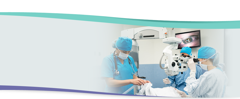
The vitreous gel which is pulling on the retina is removed from the eye and usually replaced with a gas bubble. Your body’s own fluid will gradually replace the gas bubble. Sometimes this procedure is combined with scleral buckle. The length of the surgery varies from one to several hours, depending on your condition. This surgery may need to be combined with cataract surgery if the lens of the eye is not clear (cataract) which makes it difficult for the surgeon to see into the eye properly. The ophthalmologist view’s your eye through a microscope. Various miniature instruments are placed into the eye through tiny incisions in the sclera (white part of your eye).
Your ophthalmologist will confer with an anaesthetic colleague to decide whether local or general anaesthesia is best for you. You may have to stay overnight in the hospital. You will need to have a physical examination to alert your ophthalmologist to any special medical risks. A painless ultrasound test maybe performed to view the inside of the eye.
You can expect some discomfort after surgery. You will need to wear an eye patch for a short period of time. Your ophthalmologist will prescribe any necessary medications for you and advise you when to resume normal activity. Flashes and floaters may continue for a while after surgery. You should take note and report this to the ophthalmologist. Improvement in your vision after surgery will depend on many variables.
DO NOT FLY IN AN AEROPLANE OR TRAVEL UP TO HIGH ALTITUDES IF YOU HAVE A GAS BUBBLE INSIDE THE EYE
A rapid increase in altitude can cause a dangerous rise in eye pressure if gas has been placed inside the eye.
After the operation the vision may take some time to improve. A change in glasses after several months may help.
Most retinal detachment surgery is successful, although a second operation is sometimes needed. If the retina cannot be reattached then the eye will continue to lose sight and ultimately become blind. Vision may take up to months to improve and in some cases may never return fully. The more severe the detachment, the less vision may return.